– Devika Rege
Extensive research which followed the discovery of DNA, to know more about its structure, function and mechanisms, led us to believe that each cell of our body contains the same DNA molecule – which is exclusive for each person. Due to this understanding, the possibility of encountering any instance of intra-organismal genetic variation was a far cry from reality; however, discoveries of mosaicism and chimerism in the early 1950s have forced us to rethink our beliefs while catalyzing ground-breaking research in the fields of evolutionary and developmental embryology.
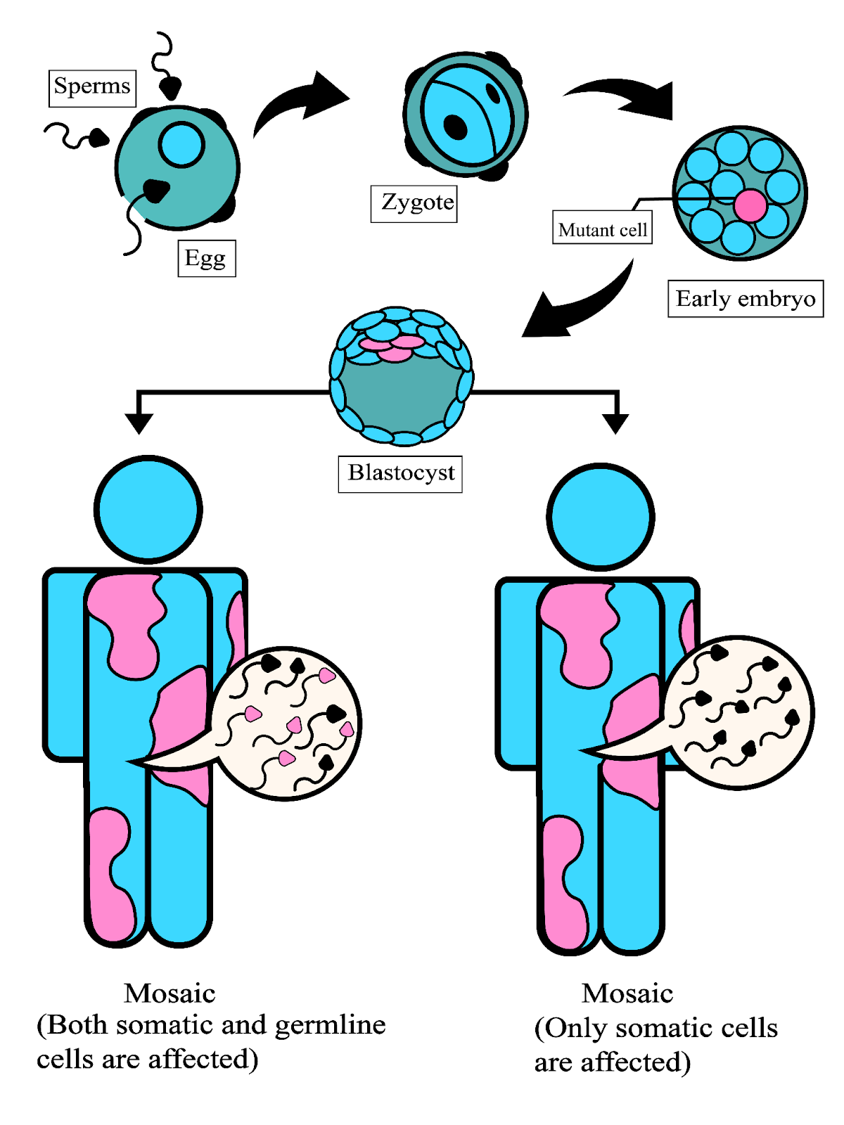
Figure 1 depicts the two different types of mosaicism [L-R]: Where both somatic and germline cells are affected and when only somatic cells are affected
Illustrated by Faria Shahreen Ahmed
Mosaicism is an intriguing phenomenon in which the presence of a few genetically distinct cells is observed within an organism. Factors such as environmental changes, sudden genetic mutations, reversal of existing mutations and chromosomal abnormalities contribute to mosaicism in the nuclear or mitochondrial DNA. If the instance resulting in mosaicism takes place during the development of the organism, then, the somatic and germline cells both are at risk of mosaicism, which – if affecting the germline, could be passed on to the next generation[Fig.1]. On the contrary, if the instance resulting in mosaicism takes place after development, then only the somatic cells might be at risk of mosaicism, with no possibility of a generational transfer [Fig.1]. The physical consequences or onset of a disorder due to mosaicism depends upon the amount and type of mosaicism present in the organism. Hereditary tyrosinemia (type I) is a type of Mendelian disorder in which the FAH gene is mutated which results in the affected individuals having a liver mosaic consisting of normal and mutant cells, which results in nodule formation in the liver and presents a growth advantage. (Chial, 2008) Bloom syndrome is another example of a single gene disorder in which the BLM gene which codes for DNA helicase is mutated, due to which supercoiling in the DNA strands during replication cannot be averted. A consequence of which is a high occurrence of recombination in sister chromatids is observed in those affected with Bloom syndrome, resulting in chromosomal instability. (Chial, 2008)
Chimerism is another phenomenon in which a person contains two different sets of DNA which are derived from two or more zygotes. (Madan, 2020) While mosaicism and chimerism may seem like two versions of the same idea, their zygotic origin makes them as distinct as two parallel lines. Chimerism could be caused due to intra-organismal genetic variations which occur during pregnancy or inter-organismal genetic variations which occur during tissue grafts or allogeneic fusions (fusion of genes from different individuals of the same species7); while the former is referred to as natural chimaeras, the latter are considered to be artificial. (Madan, 2020) Also referred to as ‘fetomaternal microchimerism’ (FMM), this phenomenon involves bidirectional transport across the placenta during pregnancy. (Hahn et al., 2019; Madan, 2020) Evidence of this was provided when male foetal leucocytes were detected in mitogen-stimulated maternal blood samples; their nature was confirmed due to the presence of the Y chromosome. (Hahn et al., 2019) While this finding confirmed previous research, which hypothesized the presence of FMM, it also paved the way for focused research which deepened our understanding of the subject. It was further reported that these foetal cells persist in the maternal body for a long time and could be detected as long as three decades post-pregnancy; this finding also opened up a new research area which involves finding out if such this persistence contributes to putting women at an increased risk of autoimmune disease post-pregnancy. (Hahn et al., 2019; Madan, 2020) Another study conducted by Holzgreve W and Hahn S (2000) reported that an increased amount of male foetal cells in the maternal blood were an early sign of developing preeclampsia – a grave condition in pregnant women which is characterized by high blood pressure and could prove to be fatal for the foetus in absence of a timely diagnosis. Hence, this positive correlation can serve as an important diagnostic feature to prevent preeclampsia even in absence of evident signs. The trends and rate of this bidirectional trafficking were also found to be an important indicator of preeclampsia with normal healthy women having a rate of 16% in the second trimester, 29% in the third trimester, and 14% post-pregnancy – these same rates or rather no micro chimerism was witnessed in women who later developed preeclampsia. (Gammill et al., 2011; Madan, 2020)

Illustrated by Froilan Sequeira
While mosaicism and chimerism both contribute to genetic variation, the extent of their contribution is significantly different with the latter having a greater influence on the resulting genetic composition. (Santelices, 2004) Apart from this, both phenomena also differ in the organisms in which they occur and their frequency of occurrence in them; while chimerism is observed to occur in plants, seaweeds, sponges, corals and ascidians in a particular type of environment, mosaicism is found to occur in all organisms including the ones mentioned above without any specific environmental requirements. (Santelices, 2004) Aside from contributing to diseases or as disease indicators along with their contrasting nature, these phenomena have contributed to our knowledge of development and origin while also providing us with guaranteed food for thought.
Did you enjoy reading this? Let us know what you thought of this article in the comments section below!
ABOUT THE AUTHOR
Devika Rege
The author is an undergraduate student studying Life Science and Biochemistry at St. Xavier’s College, Mumbai.
We would like to thank Dr. Surabhi Sonam , D Y Patil International University, Akurdi, Pune for reviewing this article and for her valuable inputs.
-The Boffin Bloggers
References
- Chial, H. (2008). Somatic Mosaicism and Chromosomal Disorders. Nature Education. https://www.nature.com/scitable/topicpage/somatic-mosaicism-and-chromosomal-disorders-867/.
- Madan, K. (2020, June 18). Natural human chimeras: A review. European Journal of Medical Genetics. https://www.sciencedirect.com/science/article/pii/S1769721220302895.
- Hahn, S., Hasler, P., Vokalova, L., van Breda, S. V., Than, N. G., Hoesli, I. M., Lapaire, O., & Rossi, S. W. (2019). Feto-Maternal Microchimerism: The Pre-eclampsia Conundrum. Frontiers in Immunology, 10. https://doi.org/10.3389/fimmu.2019.00659
- Hahn, S., & Holzgreve, W. (2002). Fetal cells and cell-free fetal DNA in maternal blood: new insights into pre-eclampsia. Human Reproduction Update, 8(6), 501–508. https://doi.org/10.1093/humupd/8.6.501
- Gammill, H. S., Adams Waldorf, K. M., Aydelotte, T. M., Lucas, J., Leisenring, W. M., Lambert, N. C., & Nelson, J. L. (2011). Pregnancy, Microchimerism, and the Maternal Grandmother. PLoS ONE, 6(8), e24101. https://doi.org/10.1371/journal.pone.0024101.
- Santelices, B. (2004, September 15). Mosaicism and chimerism as components of intraorganismal genetic heterogeneity. Wiley Online Library. https://onlinelibrary.wiley.com/doi/full/10.1111/j.1420-9101.2004.00813.x.
- Medicinenet.com. (2021). Retrieved 11 August 2021, from https://www.medicinenet.com/allogeneic_graft/definition.htm.
